UCT researchers discover bone disease in a 265 million-year-old mammal ancestor


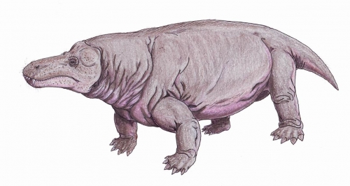
Reconstruction of Jonkeria: Wikipedia
Researchers at the University of Cape Town’s Biological Sciences Department have discovered an unusual bone tissue pattern that was suspected to be osteomyelitis in the femur of an omnivorous therapsid, more specifically known as a dinocephalian.
Osteomyelitis is a degenerative bone disease caused by a bacterial infection which eats away bone. It is common in modern mammals and reptiles as well as in their earliest prehistoric ancestors, which predated the dinosaurs.

Dr Chris Shelton (left) with Professor Anusuya Chinsamy-Turan (right)
Dr Christen Shelton, Department of Biological Science and first author of the paper published in the International Journal of Paleobiology, Historical Biology, said: “While analysing thin sections of the femur under a microscope, I noticed that the bone tissue did not follow the normal growth pattern as that observed in other specimens.”
The earliest occurrence of this disease was discovered in the backbone of the iconic dorsal sailed pelycosaur. This “mammal-like reptile” lived 280 million years ago and was part of a group that gave rise to the mammals we know today. Linking modern mammals and pelycosaurs is another group of advanced “mammal-like reptiles” known as the therapsids. Until now osteomyelitis was assumed to exist in this group of mammal-like reptiles as well, but no proof had ever been found.
Shelton suspected this was something special and sought expert advice from Professor Anusuya Chinsamy, a palaeobiologist at UCT. She concurred that this was indeed unusual, pointing out areas of the bone tissue which showed some bone layers growing perpendicular to each other. Chinsamy noted that: “This pattern is often indicative of a pathology, something that either damaged or disrupted the normal bone tissue growth.”

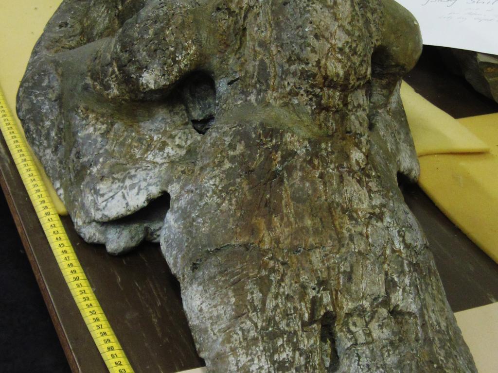
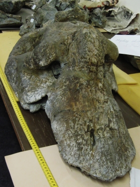
Skull of Jonkeria specimen from collections at ESI, Wits University
Bruce Rothschild, M.D. of the West Virginia University School of Medicine, who has worked extensively researching bone pathologies in dinosaurs, joined the team of researchers and identified that: “The unusual bone pattern was in fact due to a bacterial infection and the animal’s response to it.”
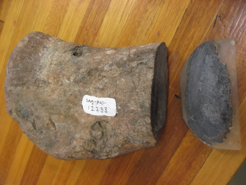
“This made sense because on the femur we found two teeth puncture marks, which we believe resulted from a bite by a predator during its lifetime. This potential bite became infected and resulted in the pathology we discovered here,” Shelton added.
Chinsamy-Turan, a global expert on the microscopic structure of the bones of extinct and extant vertebrates, noted that: “Researchers have long assumed that osteomyelitis must have occurred in therapsids, but now we can back up those assumptions with histological proof.”

Jonkeria femur sampled. Note the two tooth puncture marks
Thanks to this research, there is now further evidence of the link between mammals and their “mammal-like” reptile ancestors.
According to the New World Encyclopedia, therapsids are "mammal-like reptiles" that flourished from the Early Permian to the Late Triassic periods (c. 275 - 205 million years ago) and are thought to have been the precursors of mammals. Aside from the mammals, all the other lines of descent from the therapsid ancestors have become extinct.
